Article
Speculum Examination
By Patrick M. McCue, DVM, PhD,
Diplomate American College of Theriogenologists
A vaginal speculum examination is a routine component of a mare reproductive evaluation. The goals of a speculum examination are to evaluate anatomic characteristics of the cervix relative to stage of the estrous cycle and to detect abnormal conditions of the vagina and cervix, such as urine pooling, inflammation, trauma, and vaginal varicose veins. The procedure takes only a few minutes to perform and uses a minimum amount of equipment.
To perform the examination, the mare's tail is wrapped and her perineum washed with mild soap and water, rinsed thoroughly and dried with paper towels. A disposable sterile vaginal speculum is lubricated and gently inserted through the vulva and into the vestibule. The speculum is advanced forward and upward until it has passed through the transverse fold into the vagina, at which point a slight sucking sound may be audible as air is aspirated into the vagina. The speculum is then advanced horizontally toward the anterior or front end of the vagina. A bright pen-light or other light source is used to illuminate the vaginal vault.
The vaginal vault often inflates with air during passage of the speculum, which facilitates visual examination. The interior of the vagina is thoroughly examined and the evaluation continues as the speculum is slowly withdrawn.
The appearance of the vaginal portion of the cervix varies with season, stage of the estrous cycle, pregnancy status, and presence of infection. The cervix of a non-cycling mare (i.e. a mare in winter anestrus) appears pale, dry and may be open. When a mare is cycling and in heat, the cervix relaxes onto the floor of the anterior vagina, becomes edematous, pink and is moistened with clear mucus. After ovulation, as progesterone levels increase, the cervix becomes pale and dry, is tightly closed, and is elevated off the floor of the vagina. The cervix of a pregnant mare is tightly closed and may be covered by a thick sticky mucus plug. In the presence of a uterine infection, the cervix becomes reddened and may have a white or cream-colored discharge coming out of the opening from the uterus.
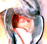
Urine pooling is recognized by an accumulation of cloudy yellow fluid in the anterior vagina. Pooling of urine is most common in older mares with poor perineal conformation. The condition occurs when urine refluxes forward into the vaginal cavity instead of exiting out of the vulva during urination. Once in the vagina, urine may pass into the uterus as the cervix relaxes during estrus, resulting in significant inflammation and a decrease in fertility.
A speculum examination may also be performed to determine the presence (or absence) of trauma to the vagina and cervix following a traumatic breeding. For example, if a stallion has blood on his penis after dismounting a mare during a live cover, a speculum examination can help determine if the source of the bleeding is the mare. Causes of bleeding following a live cover include perforation of a hymen in a maiden mare, disruption of a vaginal varicose vein, and trauma to the vagina and/or cervix. The most serious of these conditions is a full-thickness tear through the anterior vaginal wall into the abdominal cavity, which would warrant immediate veterinary medical care.
The hymen is located at the junction of the vestibule and vagina, approximately 3 to 4 inches inside the vulva. The hymen may be complete, occluding the entire vaginal opening, or may be a partial membrane. Tearing of the hymen during first breeding or insemination results in disruption of small blood vessels that traverse the membrane and transient minor bleeding.
One or more varicose veins may be present on the lateral wall near the junction of the vestibule and vagina. These blood vessels may spontaneously bleed or may be disrupted during live cover or insemination procedures. In most instances the bleeding will stop without treatment and the amount of blood lost is not clinically significant.
In summary, a vaginal speculum examination can provide valuable diagnostic information that may be readily obtained by other techniques. Please consult with your equine veterinarian for more information on breeding soundness evaluations.
Figure 1. Urine pooling in the cranial vagina visible during speculum examination.
.
About the Author...
Patrick M. McCue, DVM, Ph.D is Diplomate American College of Theriogenologists

Animal Reproduction Systems
800-300-5143


